"Success is the SUM of all the hard work you have put into something." -Mars
Science is NEVER resting, even if it is summer.
In the Book 5 Steps to get a 5 complete the following:
1. On a piece of paper identify the 4 Big Ideas of Biology in detail.
2. In short explain what you need to know about AP Bio exam.
3. Plan your time, use one of the outlines (or one you develop) and turn in a copy to me on the first day of school.
4. Take the diagnostic test, turn in your answer key and result into my on the first day of school.
5. For the question you were marked wrong on the diagnostic test give the correct response and WHY.
6. Elaborate on chapter 4's 'How to approach each question type.'
On the First day of school the following is due:
Read the material provided below.
Complete all virtual labs with a print out of the conclusions
Outline notes for each of the Units (don't forget diagrams).
Unit 1: AP Biology Review
Visit the Virtual Lab: Using a microscope to identify microbes
http://virtuallab.nmsu.edu/micro.php
Fundamental Biology Skills and Knowledge Overview
Chemistry, Biology and other sciences often overlap information in one form or another. A strong foundation of Chemistry knowledge is essential to understanding biology at the molecular level.
Matter
All organisms on Earth are made of matter.
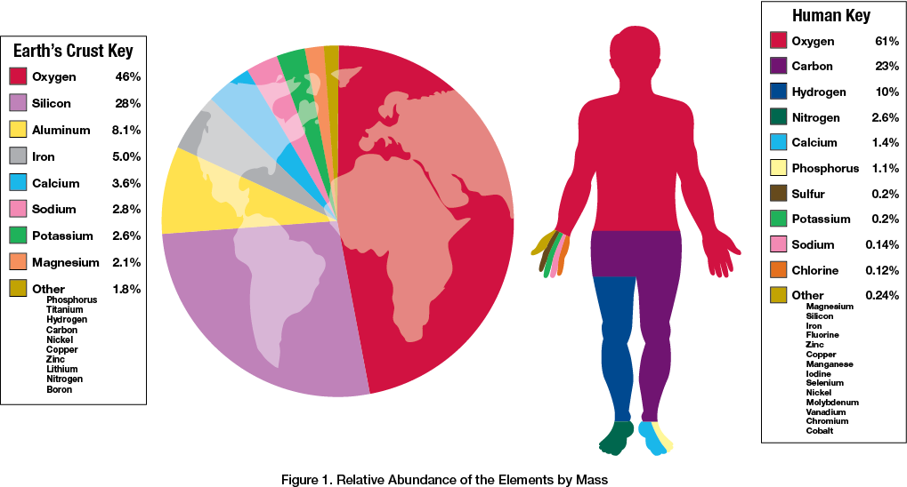
Matter is defined as any substance that takes up space and has mass. Matter is made up of atoms of elements.
An element is a pure substance that cannot be broken down to a simpler substance by chemical means. Examples of elements include hydrogen (H), copper (Cu) and oxygen (O).
A compound contains two or more different elements in a fixed ratio. Examples include Potassium chloride (KCl) and Carbon dioxide (CO2).
In Figure 1 you can see how the human body and the Earth's crust contain many of the same elements but in different proportions.
Water Is Essential
Water covers about 71% of the Earth and is essential for life. A water molecule consists of two hydrogen atoms and one oxygen atom connected by covalent bonds. See Figure 2.
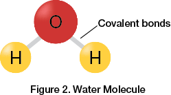
A covalent bond occurs when one atom shares electrons with another atom. Molecules held together by covalent bonds can be polar or nonpolar depending on the nature of the atoms in the bonds and the symmetry of the molecule.
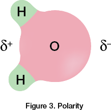
For example, water is a polar molecule. This means that one end of a water molecule has a partial negative charge and the other end has a partial positive charge. The partial charges are a result of each atom's individual electronegativity value, which is simply a number that reflects the ability of an atom to attract electrons in a covalent bond. A more electronegative element will be more likely to attract the shared electrons. In the case of water, oxygen is more electronegative than hydrogen, so the electrons are not equally distributed between the atoms in the bonds. This creates a difference of charge across the molecule. See Figure 3.
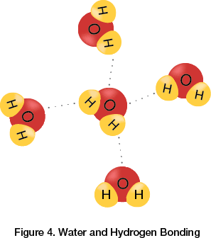
Polar molecules attract or interact more strongly with other polar molecules due to the attraction of unlike charges. The positive end of a water molecule is attracted to the negative end of a different water molecule. When water molecules interact with each other, they form hydrogen bonds. See Figure 4. Hydrogen bonds contribute to the essential properties of water and its important role in the biochemistry of living organisms.
Cohesion and Adhesion
Hydrogen bonding allows water to have unique properties, such as cohesion and adhesion. Cohesion refers to water's ability to form hydrogen bonds with other water molecules, while adhesion refers to water's ability to form hydrogen bonds with other substances.
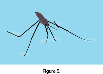
Both of these properties contribute to the high surface tension of water. Water molecules on the surface of water experience a pull from all the water molecules below the surface due to cohesion. For example, the water molecules at the top of the water in a beverage glass do not have water molecules surrounding them from every angle, so they cling more closely to the water molecules they are touching. Surface tension is what allows bugs to walk on water.
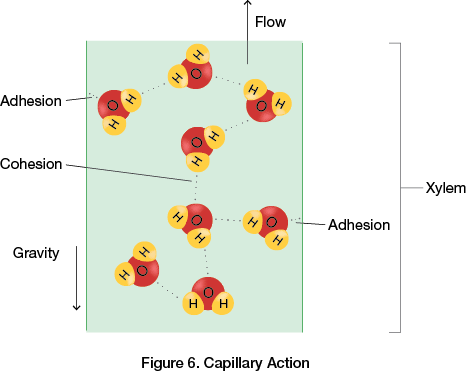
Cohesion, adhesion and surface tension are responsible for capillary action in plants.
Capillary action is the process by which a plant takes up water against gravity without using energy.
Cohesion holds water molecules together as they are pulled up through the plant to replace water that has been lost through the leaves.
Adhesive forces between the water molecules and the hydrophilic walls of the xylem allow the water to move against gravity. See Figure 6.
Density of Water
When water is in a solid state, water molecules are arranged in a crystal formation. The hydrogen bonds in the crystal formation of ice are locked, whereas the hydrogen bonds in liquid water are continuously breaking and reforming, which allows the liquid water molecules to be closer together. Because of the rigid crystal formation, ice has fewer water molecules per a given volume of water, resulting in a lower density than liquid water.
When ice and water are together, the ice will float because it is less dense. For example, in the winter, a lake or pond may have a frozen layer of ice on top, which allows life below the ice to survive beneath the frozen surface. This is a unique property of water since, in general, solids are usually more dense than liquids. However, liquid water is more dense than ice.
The Universal Solvent
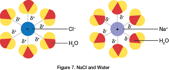
Water is often referred to as the universal solvent because many substances are able to dissolve in water due to the polar nature of water molecules. Substances that are hydrophilic have an affinity or are attracted to water. Ionic solids are hydrophilic and readily break apart when mixed with water. For example, NaCl, sodium chloride, breaks apart into Na+ and Cl? ions when added to water. The positive ends of the water molecules, the hydrogens, surround the negative chlorine ion, while the negative ends of the water molecules, the oxygens, surround the positive sodium ion. See Figure 7.
Although it is called the universal solvent, water does not actually dissolve every substance. Hydrophobic substances are not attracted to water because they are nonpolar. Most living tissue is surrounded by a hydrophobic layer or membrane to protect tisues from dissolving in water!
pH and Living Organisms
As mentioned previously, water is composed of two hydrogen atoms and one oxygen atom. Sometimes a hydrogen atom that is part of a water molecule will leave its original molecule and join a different water molecule. When that happens, a hydrogen ion (H+) is transferred from one molecule to another. The hydrogen ion is a single proton with a 1+ charge. The water molecule that lost a proton is now a hydroxide ion (OH?) and the water molecule that gained a proton is now a hydronium ion (H3O+). A hydronium ion is also referred to as an H+ ion.
This property of water is important in biology as H+ and OH? are very reactive ions. Changes in their concentration can drastically alter cellular proteins and other molecules. Furthermore, changes in concentration of these ions are measured by the pH scale. See Figure 8.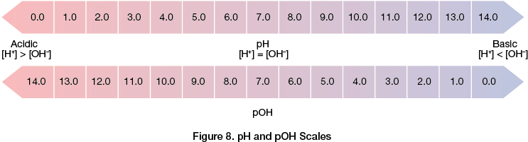
The concentration of H+ ions is inversely related to the pH value. pH is the negative log of the hydrogen ion concentration:
A neutral aqueous solution has an H+ concentration of 107 and an equal concentration of OH ions. To determine the pH value:
Notice that as the pH value declines, the hydrogen ion concentration increases. By knowing the hydrogen ion concentration, the hydroxide ion concentration can also be determined. The pH scale goes from 0 to 14. If the solution has a pH of 4, the hydrogen ion concentration, [H+], is 104, which implies the hydroxide ion concentration [OH] is 1010 according to the equation:
A buffer is a solution containing a weak acid and its corresponding base. Buffers are essential in biological fluids because their role is to resist changes in pH when acids or bases are added.
Carbon
Living matter on Earth primarily consists of Carbon, Oxygen, Hydrogen and Nitrogen. There are also small amounts of sulfur and phosphorus in biological molecules. Lipids, proteins, DNA and carbohydrates are present in living matter. These large molecules are comprised primarily of carbon. Biological diversity at the molecular level results from carbons ability to form numerous molecules with different shapes and chemical properties.
Carbon is capable of bonding to many other atoms such as oxygen, hydrogen and nitrogen. Carbon can also bond to other carbon atoms to form long organic carbon chains. Chains consisting of only carbon and hydrogen are known as hydrocarbons. Hydrocarbon chains in lipids (fats) store energy for later use.
Important Biological Functional Groups
Functional groups can replace one or more of the hydrogens bonded to the carbon skeleton of a hydrocarbon to create different types of organic molecules. These functional groups participate in chemical reactions and sometimes contribute to function by affecting the shape of a molecule. See Figure 9 for a table describing common functional groups.
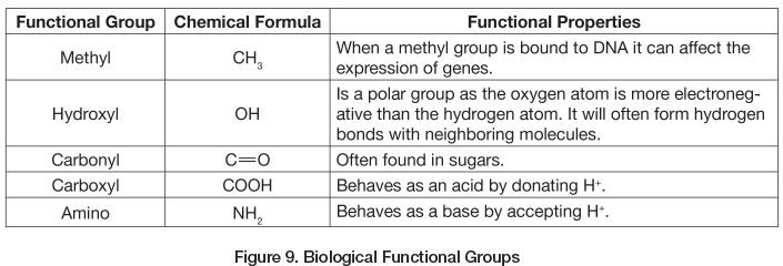
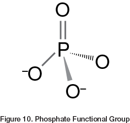
All functional groups, with the exception of the methyl group, increase the solubility of a molecule. The phosphate functional group deserves special mention as it is extremely important in many cellular processes. This ionized group contains a phosphorus atom bonded to four oxygen atoms. See Figure 10. The phosphate group is important because molecules with phosphate groups are able to react with water to release energy.
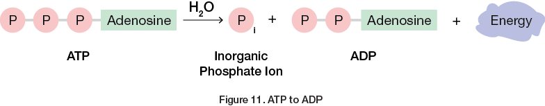
Adenosine triphosphate, ATP, is a type of organic phosphate and the primary energy-transferring molecule in cells. It consists of three phosphate groups with adenosine attached to one end. When ATP reacts with water, it forms an inorganic phosphate and ADP, adenosine diphosphate. This chemical reaction releases energy that is used by the cell. See Figure 11.
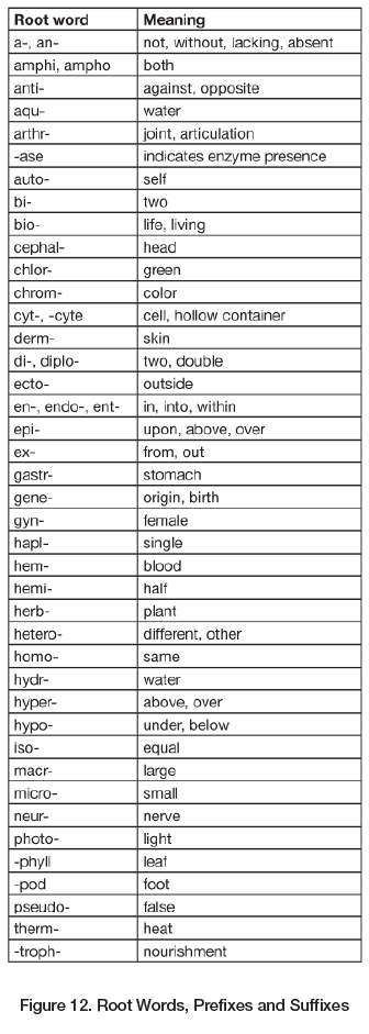
Root Words, Prefixes and Suffixes
Biology is full of specific terminology, but there are many common root words, prefixes and suffixes. Many words in the English language are formed by taking basic words and adding different prefixes and suffixes to them. The basic words are known as root words. By learning common prefixes and suffixes it becomes easier to decipher meanings of new, unfamiliar words. Figure 12 is a chart of the most common root words, prefixes and suffixes used in biological sciences.
Summary
- An element is a pure substance that cannot be broken down to other substances. A compound contains two or more different elements in a fixed ratio.
- The hydrogen and oxygen atoms of an individual water molecule are connected via covalent bonds. Water molecules bond with each other via hydrogen bonds.
- Water is polar. The oxygen carries a partially negative charge and the hydrogen carries a partially positive charge.
- Water's cohesive and adhesive properties, density, and ability to act as a universal solvent all contribute to life on Earth.
- pH is an expression of [H+]. Hydrogen ion concentration varies because of water's ability to form hydronium and hydroxide ions, which interact with other ions dissolved in water.
- Functional groups can replace a bonded hydrogen in a hydrocarbon to create different types of organic molecules.
- Molecules with phosphate groups are able to react with water.
********************************************************************************************************************************************************************************
AP Biology Review Unit 2
Virtual Lab: http://bio.rutgers.edu/~gb101/lab1_cell_structure/section8_frames.htmlhttp://bio.rutgers.edu/~gb101/lab1_cell_structure/section8_frames.htmlUnit
Overview: Cells Structure and Function
http://www.cellsalive.com/cells/3dcell.htm
The goal of this unit is to understand that cells are responsible for essential life functions such as energy transfer and transformation, gas exchange, waste disposal, growth, reproduction and environmental interaction. Cells contain internal structures that carry out specialized life functions.
Prokaryotic and Eukaryotic Cells Similarities and Differences
An interactive Model of the Cell:
http://www.nsf.gov/news/overviews/biology/interactive.jsp
Cells are classified as either prokaryotic or eukaryotic. Both prokaryotic and eukaryotic cells share the following characteristics: both are enclosed by a plasma (cellular) membrane and contain DNA and ribosomes. The plasma membrane serves as a selective barrier between the components of the cell and its surrounding environment. The DNA, while different in structure between the two cell types, carries the cell's genetic information on chromosomes. The ribosomes are essential for protein synthesis.
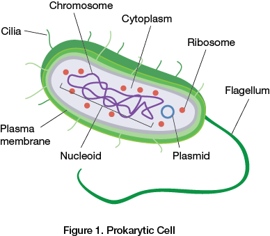
Prokaryotic cells are microorganisms such as bacteria and archaea. They are smaller than eukaryotic cells with a diameter of 0.1 to 5 nm. Prokaryotes also lack a membrane-bound nucleus. Instead, the chromosome is concentrated in a region known as the nucleoid. Many bacteria also have circular DNA molecules called plasmids, which replicate separately from the other chromosome. Prokaryotes may have a flagellum, which is a long appendage used for locomotion. Cilia may also be present and is used to control a cell's movement.
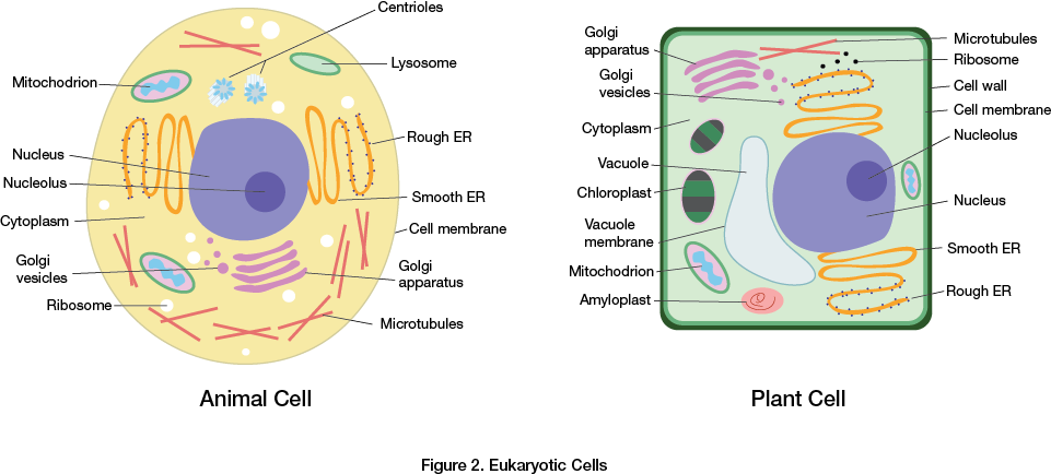
Eukaryotic cells include plant and animal cells. The DNA of a eukaryotic cell is found in the nucleus. It is surrounded by a double membrane called the nuclear envelope. The DNA is in linear form and condenses into tightly-packed chromosomes prior to replication.
Eukaryotic cells are also filled with cytoplasm; a thick solution consisting of water, salts and proteins. Cytoplasm is located between the nuclear membrane and the plasma membrane. Eukaryotes contain a variety of organelles throughout the cytoplasm. (Organelles are not present in prokaryotic cells.) Eukaryotes are much larger than prokaryotes with an average diameter of 10-100 micrometers
Plant cells and animal cells are types of eukaryotic cells.
Eukaryotic Organelles A Detailed Look
Eukaryotes have a variety of different organelles, each with a specialized structure and function.
Check out this website for Cell Structure:
http://www.nigms.nih.gov/Education/pages/FactSheet_Cells.aspx
The endomembrane system consists of the nuclear envelope, endoplasmic reticulum, Golgi apparatus, lysosomes, vesicles, vacuoles and the plasma membrane. The purpose of this system is to allow cells to exchange, process and transport materials throughout the cell.
The endoplasmic reticulum (ER) consists of folded, connected, membrane-enclosed sacs called cisternae. There are two types of endoplasmic reticulum rough and smooth. The functions of theSmooth ER are different depending on the cell's type. The smooth ER can synthesize lipids, store calcium ions, process carbohydrates and also remove toxins. The Rough ER has attached ribosomes while the smooth ER does not. The function of the ER is to fold/synthesize proteins (that are produced by the ribosomes) into sacs called transport vesicles, which end up at the Golgi apparatus.
After being processed or finished in the Golgi apparatus, proteins and other products of the ER are again packaged into transport vesicles. These vesicles then travel to their final destination. For example, a secretory protein will be transported to the plasma membrane. The Golgi apparatus can also manufacture macromolecules, such as polysaccharides.
The endomembrane system in animal cells also includes lysosomes. Lysosomes are sacs of hydrolytic enzymes that digest macromolecules. Macromolecules consist of food, cellular macromolecules and damaged organelles.
Cells use specialized organelles, called mitochondria and chloroplasts, to convert energy into a usable form. Mitochondria perform cellular respiration, which is the metabolic process that uses oxygen to produce ATP. In plant and algae cells, Chloroplasts are the site of photosynthesis. Photosynthesis is the process by which light energy is converted to chemical energy.
The genetic instructions for a eukaryotic cell are found in the nucleus. The nucleus is surrounded by the nuclear envelope, which is connected to the rough ER and perforated by nuclear pores, which regulate the movement of molecules into and out of the nucleus. The nucleus stores chromosomes, which are structures made of chromatin (DNA and proteins). A nucleus can contain one or more nuclei, regions within the nucelus that are responsible for the producton of ribosomes.
Ribosomes consist of two subunits: a small ribosomal subunit and a large ribosomal subunit. Each subunit is comprised of ribosomal RNA and proteins. Ribosomes can be free in the cytoplasm or bound to the endoplasmic reticulum or nuclear envelope.
![Ribosome structure]](https://www.flinnprep.com/Assets/images/Cells_Figure_3A.png)
Cytoskeleton
Cells have a cytoskeleton, which is a network of fibers extending throughout the cytoplasm that consists of microtubules, microfilaments and intermediate filaments. Microtubules shape the cell, guide organelle movement and separate the chromosomes of dividing cells. They are the largest component of the cytoskeleton at 25 nm in diameter. In animal cells, microtubules provide strength to the cytoskeleton.
Microfilaments are 7 nm in diameter and consist of two intertwined strands. The primary role of microfilaments is to bear tension. They also aid in maintaining cell shape, muscle contraction, cytoplasmic streaming, cell motility and cell division. Cytoplasmic streaming is the flow of cytoplasm throughout the cell in order to distribute materials evenly.
Intermediate filaments are fibrous proteins supercoiled into thicker cables. They are typically 8-12 nm in diameter and function to maintain cell shape, anchor the nucleus and other organelles, and help form the nuclear lamina, which provides structure to the nuclear envelope. See Figure 4.
Cellular Membranes Phospholipid Bilayer
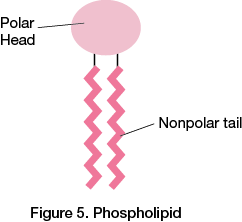
The plasma membrane functions as a selective barrier that allows oxygen, nutrients and wastes to pass into and out of the cell. The plasma membrane is a phospholipid bilayer, meaning it has two layers of phospholipids. Phospholipids are amphipathic in that they have a hydrophobic (water-fearing) region and a hydrophilic (water-loving) region. The head of a phospholipid, the hydrophilic region, contains a phosphate group, which makes it polar. The hydrophobic tail contains fatty acid hydrocarbon chains, which are nonpolar. See Figure 5.
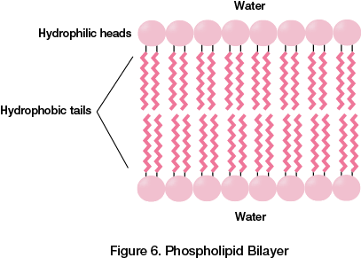
When phospholipids are placed into a solution, the hydrophobic tails will orient themselves facing inwards and toward each other, while the hydrophilic heads will be exposed to the water area. Hence, a lipid bilayer is formed. See Figure 6.
Cells are constantly exchanging molecules and ions across their membranes. This process is regulated by the selective permeability of the plasma membrane. Nonpolar substances are soluble in the lipid bilayer and easily pass through. Conversely, polar molecules and ions generally cannot pass through the lipid bilayer without the aid of transport proteins.
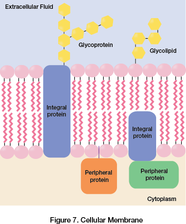
Cellular Membranes Membrane Proteins
Phospholipids and proteins are arranged in cell membranes according to the fluid mosaic model.The membrane itself is a fluid structure containing various proteins embedded in or attached to the phospholipid bilayer. The unsaturated hydrocarbon tails of the phospholipid bilayer are kinked, which prevents them from packing tightly together and therefore ensuring membrane fluidity. It is important to note that plant cell walls have a different structure composed of cellulose fibers embedded in other polysaccharides and proteins.
There are two main types of membrane proteins: integral proteins and peripheral proteins.Integral proteins are embedded in the lipid bilayer. Peripheral proteins are attached to the membrane surface and not embedded in the lipid bilayer at all. The purpose of membrane proteins is to aid in transport, enzymatic activity, signal transduction, cell to cell recognition, intercellular joining, and attachment to the cytoskeleton and ECM. Animal cells secrete glycoproteins and glycolipids, which are short chains of sugars on the exterior side of the plasma membrane that are linked to proteins or lipids and interact with the surface molecules of other cells to form the extracellular matrix (ECM). See Figure 7.
Passive Transport
The primary rule of diffusion is that in the absence of other forces, molecules or particles willmove from a region of higher concentration to a region of lower concentration. This is known as moving down its concentration gradient. When diffusion occurs across a biological membrane, such as a cell membrane, this is called passive transport. Passive transport is the diffusion of a substance through a biological membrane with no expenditure of energy.
Osmosis is an example of passive transport. Osmosis is the diffusion of water across a selectively permeable membrane. Water will flow from an area of lower solute concentration (called a hypotonic solution) to an area of higher solute concentration (hypertonic solution). If two solutions have an equal solute concentration, they are considered isotonic. Water molecules move through specialized channels called aquaporins.
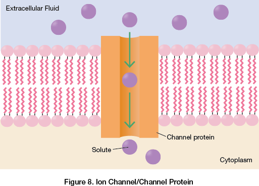
Facilitated diffusion is a type of passive transport that occurs when a transport protein increases the speed at which a solute moves across a membrane down its concentration gradient. An ion channel assists in the diffusion of ions across a membrane using a channel protein. See Figure 8.
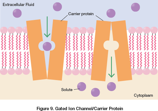
Gated ion channels use carrier proteins that open and close in response to a stimuli. See Figure 9. Even though facilitated diffusion uses transport proteins, it is still considered to be passive transport because the solutes are moving down a concentration gradient.
Active Transport
When a solute moves across a biological membrane against a concentration gradient (from an area of low concentration to high concentration), it requires energy. This is called active transport. Active transport uses carrier proteins and energy from ATP to move a solute against the concentration gradient. One common process in a cell that uses active transport is the sodium/potassium pump.
Exocytosis & Endocytosis
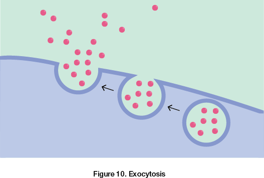
Large molecules require packaging in vesicles to cross the plasma membrane. This type of bulk transport requires energy. Exocytosis is the transport of large substances produced inside the cell to the outside of the cell. The cell encloses a substance in a vesicle that fuses with the plasma membrane. The plasma membrane evacuates the contents of the vesicle to the interior of the cell. See Figure 10.
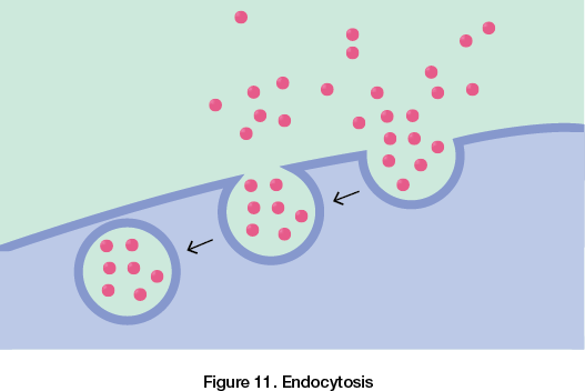
Endocytosis is basically the reverse of exocytosis. The cell takes in substances by forming new vesicles in the plasma membrane. A region of the plasma membrane will cave inwards toward the center of the cell. As the pocket forms, it pinches off the plasma membrane releasing the vesicle into the cell.
Cellular Communication
Communication among cells is continuous. Cell junctions connect adjacent plant and animal cells. Plant cells have plasmodesmata, channels that pass through adjoining cell walls connecting chemical environments to allow water and small solutes to move freely between the cells. Animal cells have tight junctions, desmosomes and gap junctions:
• Tight junctions prevent leakage of material through the space between cells by using proteins to bind the membranes tightly to each other.
• Desmosomes function as rivets, fastening cells together.
• Gap junctions consist of proteins that surround a pore and allow the passage of materials between cells.
Neurons and Cell Signaling
When a person touches something very hot, they remove their hand as quickly as possible. This is possible because the body uses the nervous system to send and receive messages from one area to another.The nervous system processes information in three stages: sensory input, integration and motor output. In this example, the sensory input would be the sense of touch that is feeling the hot item, integration is how the central nervous system processes that message, and motor output signals the body to immediately remove the hand from the hot surface.
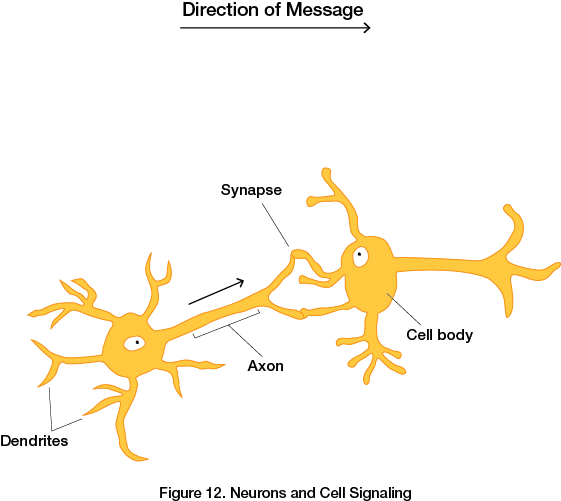
The central nervous system (CNS) consists of the brain and spinal cord. The peripheral nervous system (PNS) is made of neurons that deliver messages to and from the central nervous system to the rest of the body. Neurons have three general regions that perform specific functions: dendrites, axons, and the cell body, which contains the nucleus and organelles. The majority of neurons have dendrites that receive information from neighboring neurons. The axon is an extension of the neuron that transmits electrical signals along its length to the synapse, which is the space between the axon of one neuron and the dendrite of the next neuron. The electrical signal triggers chemicals called neurotransmitters to move across the synapse and communicate the message to the next neuron, which in turn activates another electrical signal.
Neurons also communicate via electrical signals. At rest, cells have a particular voltage. When a message is sent via electrical signal, gated ion channels open and close to allow ions to enter and exit the cell. This increases the voltage and transmits the message.
Relationship of Surface Area to Volume
Why are cells so small?
Each region of a cells’ surface can only pass a limited amount of substances through it per second. Therefore, the ratio of surface area to volume is extremely important. Smaller cells have a greater ratio of surface area to volume than larger cells. This is because as an object increases in size, its volume increases at a greater rate than its surface area. Cells need a surface area that is large enough to accommodate their volume. This is why living organisms have many small cells as opposed to fewer large cells.
Summary
- Prokaryotic and eukaryotic cells are different. Prokaryotic cells lack nuclei and membrane-bound organelles. Eukaryotic cells have organelles that perform specific functions.
- Some organelles are found in both plant and animal cells while others are exclusive to one or the other.
- In eukaryotes, genetic instructions are found in the nucleus.
- The endomembrane system consists of the nucleus, endoplasmic reticulum, Golgi apparatus, lysosomes, vesicles, vacuoles and plasma membrane. Its function is to process proteins, perform metabolic functions, and provide transportation of materials.
- Mitochondria (animal and plant cells) and chloroplasts (plant cells) convert energy from one form to another.
- The purpose of a cytoskeleton is to provide structural support and assist in motility.
- Cellular membranes are considered fluid mosaics. Amphipathic proteins, consisting of integral proteins, peripheral proteins and glycoproteins, are embedded within the phospholipid bilayer of the membrane.
- Cellular membranes are selectively permeable.
- Passive transport is the diffusion of molecules from high to low concentrations across a membrane and does not require energy.
- Active transport requires energy to move solutes against a concentration gradient across a membrane.
- Larger volumes or molecules are transported across the plasma membrane by exocytosis and endocytosis.
- Cells are small because objects with a high surface area to volume ratio are more efficient at moving molecules (or solutes).
- Neurons transmit sensory messages to the central nervous system where they are integrated and motor responses are transmitted back to the peripheral nervous system.
- Neurons transmit signals both chemically and electrically.
**Crossword Puzzle on the cell: Fun!!!
http://publications.nigms.nih.gov/insidethecell/index_cw.html
**********************************************************************************************************************************************************************************************
AP Biology Review Unit 3
The Cell Cycle
Overview
The goal of this unit is to understand that cells of multicellular organisms continuously divide to make more cells for growth and repair. Furthermore, in multicellular organisms, the single celled zygote will continue to divide and differentiate into specialized cells that form different tissues and organs.
Cell Cycle Phases
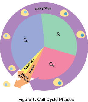
Dividing cells are always in one of two phases: the mitotic phase or interphase. The mitotic phase (M phase) consists of mitosis and cytokinesis. Interphase consists of G1, S and G2 phases. In Figure 1 below, you can see that the mitotic phase (mitosis and cytokinesis) is relatively short compared to interphase. Cells spend approximately 90% of their time in interphase.
The process of mitosis copies and distributes DNA of a cell into two identical daughter cells. Cytokinesis is the division of the cytoplasm amongst the two daughter cells.
The G1 phase of interphase stands for “gap 1.” During the G1 phase, the cell grows and makes proteins that are necessary for DNA replication. During the S phase, also known as the synthesis phase, the chromosomes duplicate. Therefore, the quantity of DNA in the cell at that time has doubled. During the G2 phase of interphase, the cell continues to grow and proteins are synthesized that are necessary for mitosis.
During mitosis, the nucleus divides and DNA is distributed into two identical daughter cells. The cytoplasm is divided between the daughter cells through a process called cytokinesis. Each daughter cell continues through the cell cycle starting back at the G1 phase.
The timing and frequency of cell division varies between different cells, tissue, organs and even species. Humans, for example, have regions where cells divide frequently such as the skin and esophagus. Conversely, in other areas of the body, such as the liver, cells can divide but only do so to repair damaged tissue. Furthermore, other cell types, such as nerve cells, never divide in a mature human. Cells that do not divide enter a phase called G0.
Chromosomes
Cell division is part of the cell cycle. The result of a cell going through the cell cycle is the production of two genetically identical daughter cells.
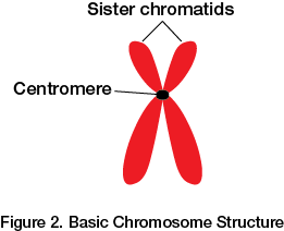
A cell’s DNA or genetic information is called its genome. In order for mitosis to occur successfully, the genetic material (DNA) must condense into structures called chromosomes. Genes are found within DNA and carry information that specify an organism’s inherited traits. The DNA and proteins that make up chromosomes are called chromatin. Each chromosome contains two identical sister chromatids. Sister chromatids are held together at the centromere. See Figure 2.
Human somatic cells (all cells except reproductive cells) contain 46 chromosomes, two sets of 23, one inherited from each parent. Reproductive cells, known as gametes, have half as many chromosomes at 23. Different eukaryotic species have a different characteristic number of chromosomes. For example, chimpanzees have 48 chromosomes per somatic cell.
Mitotic Phase
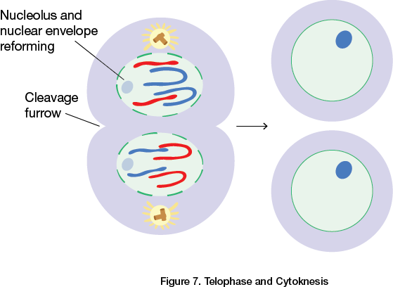
The mitotic phase includes mitosis and cytokinesis. Mitosis is the cell cycle process during which duplicated chromosomes separate into two identical daughter cells. It is characterized by five stages: prophase, prometaphase, metaphase, anaphase and telophase. Cytokinesis is a division of the cytoplasm and follows mitosis. Animal cells perform cytokinesis by cleavage and plant cells form a cell plate.
The Five Stages of Mitosis
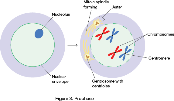
- Prophase is the first stage of mitosis. The chromatin condenses into discrete chromosomes and the nucleolus disappears. The mitotic spindle begins to form as mictrotubules extend from a region called the centrosome. The centrosome contains centrioles, but these are not necessary for the spindle to form. In fact, plant cells do not have centrioles, but still form a mitotic spindle. The short microtubules on the nonspindle side of the centrosome are called asters.
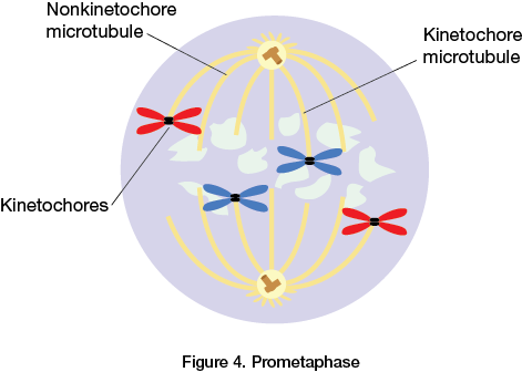
- Prometaphase is the second stage of mitosis. The nuclear envelope breaks apart and the spindle microtubules attach to a group of proteins associated with the centromere of each chromosome called a kinetochore. Each chromosome has two kinetochores. When a microtubule attaches to a kinetochore, it pulls the chromosome towards that microtubule’s pole. At the same time, a microtubule from the opposite pole attaches to the other kinetochore of the same chromosome, causing a pull in the opposite direction.
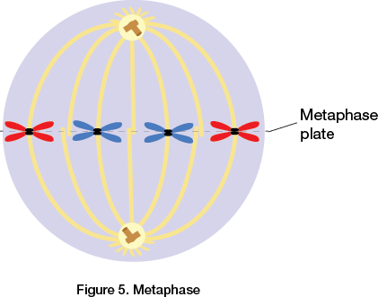
- Metaphase is the third stage of mitosis. The spindle is complete and the chromosomes that are attached to the microtubules by their kinetochores align at the metaphase plate due to the “tug-of-war” described above. At this time, the asters extend to the plasma membrane.
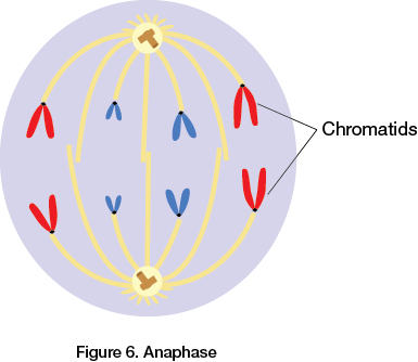
- Anaphase is the fourth stage of mitosis. The proteins holding the sister chromatids together deactivate allowing each chromosome to separate. The resulting daughter chromosomes begin "walking" towards opposite poles of the cell and the kinetochore microtubules shorten. At the same time, the nonkinetochore microtubules reduce the overlap between them to elongate the entire cell.
- Telophase is the final (fifth) stage of mitosis. The daughter nuclei form, the daughter chromosomes uncoil, and cytokinesis begins. Animal cells pinch at the cleavage furrow to separate into daughter cells. Plant cells develop a cell plate that fuses with the cell membrane to become a new cell wall between the daughter cells.
Prokaryotes and Cell Division Evolution
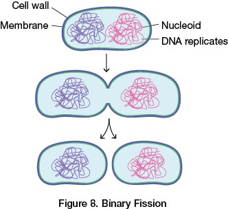
Eukaryotes divide by mitosis, but prokaryotes, such as bacteria, divide using a process called binary fission. In binary fission, chromosomes replicate and move to opposite sides of the cell. Then the cell separates into two identical daughter cells. Binary fission also occurs in single-celled eukaryotes, such as Amoeba.
There is evidence that prokaryotes existed on Earth over a billion years before eukaryotes. It is plausible that mitosis evolved from the mechanisms of prokaryotic cell division.
Cell Cycle Control Systems
It is necessary for cells to have checkpoints throughout the cell cycle to ensure that cells are replicating and dividing properly. If cells do not replicate and divide properly, then the growth and function of a cell may be adversely affected. For example, if the proteins that regulate cell division are not functioning properly, the result could be the replication and division of cancer cells.
The cell cycle is controlled by specific signaling molecules found in the cytoplasm. These molecules verify that necessary steps in the cell cycle have been accurately completed before the cell continues to the next step of the cycle.
There are three checkpoints of the cell cycle control system: G1, G2 and M. In mammals, the first and most important checkpoint is the G1 checkpoint. If a cell makes it through the G1 checkpoint, it will likely complete the subsequent stages and divide. If it does not, it will exit the cycle and remain in the G0 phase.
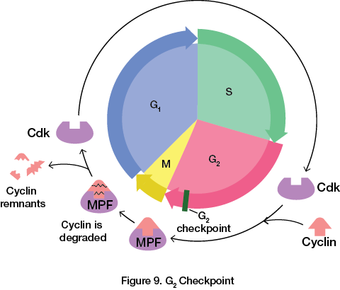
If the conditions are right, cyclin-dependent protein kinases (Cdks) allow a cell to proceed through the G1 and G2 checkpoints. Cdks are present at constant concentrations and spend a majority of the time in an inactive form. To be active, Cdks must be attached to a cyclin. A cyclin is a protein that is present in fluctuating concentrations depending on the time of the cell cycle. Cyclin-dependent kinases require cyclin to function. Their activity level fluctuates based on the cyclin concentration in the cell. Several types of Cdk-cyclin complexes form during the cell cycle. One example is the complex called thematuration-promoting factor (MPF). MPF is formed when the cyclin level rises during the S and G2 phase, and then abruptly decreases during the M phase. MPF triggers the cell to proceed past the G2checkpoint initiating and regulating mitosis. See Figure 8.
The M checkpoint is the final checkpoint in the cell cycle. In order to pass the M checkpoint, all chromosome kinetochores must be attached to the spindle at the metaphase plate. Once attached, enzymes cause the sister chromatids to separate. This results in daughter cells with the appropriate number of chromosomes.
The Loss of Cell Cycle Control
Each checkpoint determines whether or not all of the components and conditions have been met for cell division to proceed. If the cell is halted at the G1 checkpoint, the cell never progresses to the synthesis phase during which the DNA is replicated. At the G2 checkpoint, any problems in the replicated DNA trigger the cell to halt in G2 until either repairs are made or apoptosis, cell death, is prompted. If an error is detected at the M checkpoint, mitosis is halted until repairs are made. If repairs are not possible or take too long at any of the checkpoints, apoptosis is triggered and the cell dies.
When these checkpoints fail and there is a rapid, uncontrolled growth of cells, cancer is a common result. Cancer cells exhibit abnormal traits and behaviors such as excessive division, unusual numbers of chromosomes, abnormal cell surfaces, detachment from neighboring cells and the extracellular matrix, and the ability to secrete molecular signals that attract a blood supply to provide a steady stream of nutrients. Detachment and a readily available supply of nutrients allow cancer cells to grow and spread to other areas, making the cancer malignant (harmful).
Normal cells require growth factors, proteins that stimulate cell division. However, cancer cells grow and divide even when growth factors are depleted or absent. Normal cells stop dividing when the cells become crowded; this is called density-dependent inhibition. Cancer cells do not exhibit density-dependent inhibition and will grow in clumping, overlapping layers of cells.
Cancer treatments are used to attack weaknesses in cancer cells. For example, radiation damages the DNA in cancer cells, which lack the ability to repair such damage. Chemotherapy treatments interfere with the cell cycle and are toxic to actively dividing cells. For example, a chemotherapy drug may freeze mitotic spindles and thus prevent mitosis from proceeding beyond metaphase.
Gene Expression and Cell Types
As a multicellular organism develops from a zygote, it grows more cells through mitosis. Those cells become specialized through cell division, cell differentiation and morphogenesis. First, the zygote goes through a series of cell divisions that result in a copious volume of cells. Next, during embryonic development, cells undergo cell differentiation where they become specialized in structure and function and are organized into tissues and organs. The process that gives an organism its shape is called morphogenesis. It is important to note that cells differ in structure and function because they express different portions of a common genome and not because they contain different genes.
Two processes direct cells to express particular genes during early development. In the first process, maternal substances within the egg’s cytoplasm (mRNA, proteins, organelles, etc.) called cytoplasmic determinates influence early development directly. Once fertilized, early mitotic divisions distribute the zygote’s cytoplasm into daughter cells. The cytoplasmic determinants are different from cell to cell because the cytoplasm divides without making additional cytoplasm. The different cytoplasmic determinants in each cell help to regulate development by regulating gene expression during cell differentiation.
The second process is called induction. During induction, gene expression is controlled through environmental signals sent from one cell to another. Signals from surrounding embryonic cells, such as contact between cell surfaces and binding of growth factors secreted by other cells, cause changes in target cells. This creates regions of differentiated cells.
Re-cap:
- There are two main phases in the cell cycle—interphase and the mitotic phase:
– Interphase consists of G1, S and G2 stages.
– The mitotic phase consists of mitosis and is followed by cytokinesis. - DNA is packaged in chromosomes. Human somatic cells have 46 chromosomes and reproductive cells (gametes) have 23.
- Mitosis consists of five stages: prophase, prometaphase, metaphase, anaphase and telophase.
- Cells must have control mechanisms to control division so damaged cells do not continue to divide. There are three main checkpoints in the cell cycle control system: G1, G2 and M.
- When cells no longer respond or follow cellular control mechanisms or checkpoints, cancer cells may develop.
- Cells differ in structure and function because they express different portions of a common genome. They do not contain different genes. When different genes are expressed, different proteins are produced.
- Cells are signaled to express genes by cytoplasmic determinates and induction.
**********************************************************************************************************************************************************************************************
AP Biology Review Unit 4
Heredity and Variation
Overview
The goal of this unit is to understand heredity and variation. We will explore how meiosis results in offspring with unique combinations of genes from their parents. In other words, offspring inherit traits from each parent but do not become an exact replica of either parent.
Heredity
Heredity is the transfer of traits from one generation to the next. Inherited traits are transmitted on genes. Each gene in an organism’s DNA is found at a specific location on a chromosome called a locus. Humans inherit one set of chromosomes from their mother and the other set from their father, which leads to offspring that differ in appearance from each other and the parents.
Sexual reproduction combines a set of genes from each parent resulting in genetically diverse offspring. Conversely, asexual reproduction results in offspring that are genetically identical to the parent by mitosis. It simply copies all of its genes to its offspring without fusion of gametes.
Haploid and Diploid Cells
A human somatic cell contains 46 chromosomes—2 sets of 23. Two chromosomes that compose a pair have the same length, centromere position, staining pattern and genes and are called homologous chromosomes. We inherit one chromosome of each pair from each parent. Therefore, of the 46 chromosomes in our somatic cells, there are two sets of 23 chromosomes. There is a maternal set of 23 chromosomes and a paternal set of 23 chromosomes, resulting in 46 total.
The number of chromosomes present in a single set is represented by n. A cell that has two chromosome sets is called a diploid cell, represented by the number 2n. Humans have a diploid number of 46 (2n = 46), which is the number of chromosomes present in our somatic cells. Conversely, gametes (reproductive cells) contain a single set of chromosomes and are known as haploid cells represented by n. In humans, the haploid number is 23 (2n = 46 so n = 23). Different species have different diploid numbers.
Human Life Cycle
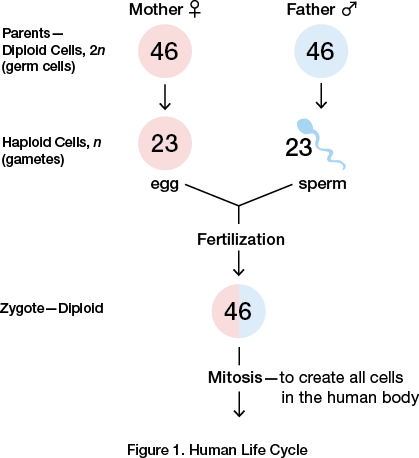
The human life cycle begins with fertilization. When a haploid sperm from the father fuses with a haploid egg from the mother, the fusion of their nuclei is called fertilization. The fertilized egg is called azygote. The zygote is diploid because it contains two sets of chromosomes. The zygote develops by mitosis to create all the somatic cells of the human body. See Figure 1 for an overview of the human life cycle process.
The only cells that are not a result of mitosis in the human body are the gametes that develop from germ cells in the gonads. Gametes are haploid and not diploid because their offspring’s zygote must contain cells with the correct number of chromosomes. If two somatic diploid cells fused to make a zygote, those cells would each contain 92 chromosomes. This result would double the number of chromosomes present in each cell in the next generation. Meiosis reduces the number of sets of chromosomes by half in the gametes to counterbalance the doubling that occurs during fertilization. This process is essential for gamete formation. Gametes are the only haploid cells in the human body and thus contain 23 chromosomes.
The human zygote has 22 pairs of autosomes and two sex chromosomes, which determine the biological sex of the offspring. A zygote with a pair of X chromosomes will be female, while a zygote with one X and one Y chromosome will be male. The Y chromosome contains the SRY gene, which controls the release of hormones. These hormones signal the development of the male during prenatal development.
Meiosis
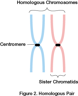
Before getting into the details of meiosis, it is important to understand some terminology. Sister chromatids are two copies of the same chromosome, which was replicated during interphase. The sister chromatids are attached by a centromere to make one duplicated chromosome. Furthermore, a homologous pair is two chromosomes—one from each parent. See Figure 2.
Unlike mitosis, meiosis has two cell divisions known as meiosis I and meiosis II that produce four haploid daughter cells. During meiosis I, the number of chromosomes is reduced from diploid to haploid. Watch the video to see what happens during meiosis.
Phases of Meiosis
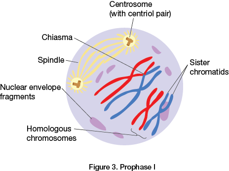
- Prophase I: Homologous chromosomes pair and exchange segments of DNA. Paired homologs become physically attached to each other by a protein structure called the synaptonemal complex. This state is known as synapsis. Crossing over is the exchange of corresponding segments of DNA between nonsister chromatids. The site of the crossover is known as the chiasma. See Figure 3.
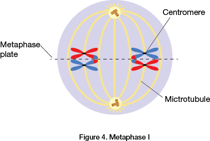
- Metaphase I: Chromosomes line up by homologous pairs on the metaphase plate. See Figure 4.
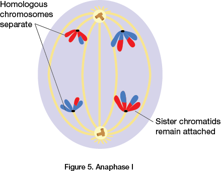
- Anaphase I: Each pair of homologous chromosomes separates towards its respective pole. Sister chromatids remain connected. See Figure 5.
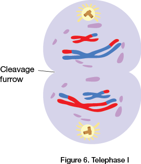
- Telophase I and Cytokinesis: During cytokinesis, the cytoplasm separates. Two haploid cells form and each chromosome contains two sister chromatids. See Figure 6.
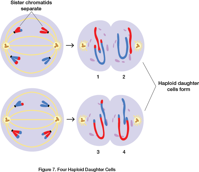
- Meiosis II: Prophase II through Cytokinesis II: Similar events occur during meiosis II. However, there is no crossing over. The main activity of meiosis II is when the sister chromatids finally separate. This produces four haploid daughter cells containing unduplicated chromosomes. See Figure 7.
Mitosis vs. Meiosis—Similarities and Differences
Both types of cell division begin with a diploid parent cell that contains a duplicated set of chromosomes. The main difference between mitosis and meiosis is that mitosis preserves the number of chromosome sets producing cells that are genetically identical to the parent. If a cell that has 46 chromosomes undergoes mitosis, the resulting cells will also have 46 chromosomes. Meiosis reduces the number of chromosomes from diploid cells to haploid cells that are genetically different from the parent. If a cell that has 46 chromosomes undergoes meiosis, the resulting cells will have 23 chromosomes. Two daughter cells are produced in mitosis while four are produced in meiosis.
Three unique events occur in meiosis I that do not occur in mitosis:
- Synapsis and crossing over occur during prophase I.
- Homologous pairs gather at the metaphase plate. In mitosis, individual chromosomes gather at the metaphase plate.
- Separation of homologs in anaphase I. Sister chromatids remain together at the centromere until they separate during meiosis II.
Chromosomal behavior during meiosis and fertilization is responsible for the majority of genetic variation. There are three main factors that contribute to genetic variation among sexually reproductive species—independent assortment, crossing over and random fertilization.
During metaphase of meiosis I, homologous chromosomes orient randomly on the metaphase plate. Each homologous pair consists of one maternal and one paternal chromosome. It is completely random whether or not a particular maternal or paternal chromosome is nearest a given pole. Therefore, there is a 50% chance a particular daughter cell will inherit the maternal chromosome and a 50% chance it will inherit a paternal chromosome. In Figure 8, you can see that the paternal chromosome of homologous pair “A” will move towards pole 1 and the maternal chromosome will move towards pole 2.
Furthermore, each pair of homologous chromosomes align on the metaphase plate independently of the other chromosomes. In Figure 8, homologous pairs A, B and C are not influenced by each other. Whether or not the paternal or maternal chromosome moves towards pole 1 in homologous set “A” will not affect the orientation of the chromosomes of homologous pairs B and C and so on. This is called the law of independent assortment.
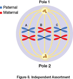
The number of possible different daughter cells is the number of chromosomes in a homolgous pair (in most cases 2) raised to the number of haploid chromosomes (n):
In Figure 8, the haploid number of chromosomes per cell is three. Since 23 = 8, there would be eight possible daughter cells.
Crossing over occurs in prophase I of meiosis I. This results in recombinant chromosomes, one chromosome carrying genes from each parent. Therefore, each chromosome in a gamete cannot be exclusively maternal or paternal. Crossing over is important because combining DNA from two parents in a single chromosome results in genetic variation in sexual life cycles.
Humans have a haploid number of 23. Therefore, the possible combinations of maternal and paternal chromosomes is 8.4 million. When taking fertilization into account, there are approximately 70 trillion diploid combinations (223 × 223). This does not account for the variations resulting from crossing over, which increases the number of possibilities exponentially. Therefore, each zygote is genetically unique.
Re-cap:
- Heredity is the transmission of traits from one generation to the next.
- Sexual reproduction combines a set of genes from two parents resulting in genetically diverse offspring.
- Two chromosomes that have the same length, centromere position, genes and staining pattern are called homologous chromosomes.
- When a haploid sperm from the father fuses with a haploid egg from the mother, the fusion of their nuclei is known as fertilization. The fertilized egg is called a zygote.
- Gametes are the only haploid cells in the human body and contain 23 chromosomes.
- Three unique events occur in meiosis I that do not occur in mitosis:
– Synapsis and crossing over during prophase I.
– Homologous pairs gather at the metaphase plate. In mitosis, individual chromosomes gather at the metaphase plate.
– Separation of homologs in anaphase I. Sister chromatids remain attached at the centromere. - Meiosis creates the genetic variation that is responsible for the development of many different traits and phenotypes that may be better suited to improve evolutionary fitness.